

168澳洲10正规开奖官网网站-开奖历史记录-开奖结果号码体彩-澳洲幸运10开奖官网直播 Our full-service HR solution delivers
We support businesses of all sizes. See how we can help you.
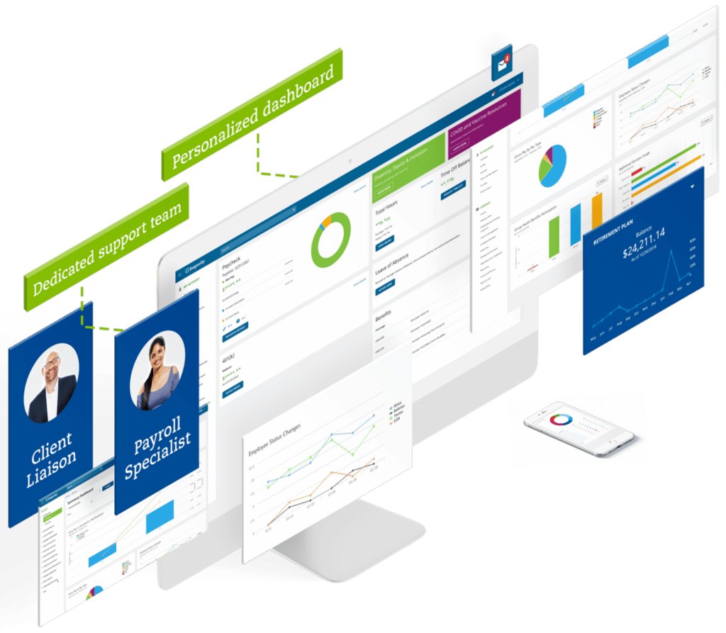
Unbeatable support meets innovative technology
Combine our unmatched service with powerful HR technology, and managing your most important asset ‒ your people ‒ gets a whole lot easier.
Explore the solution that fits your company.
10 reasons why you’ll love our HR technology
Spending more time bogged down in frustrating HR tasks and less on your business? Our human capital management platform eases your burden and supports you and your employees every step of the way.

2023年澳洲幸运10官方最新开奖 168澳洲幸运10官网历史查询 :查询开奖结果APP 查询开奖记录 See how Insperity customers are finding success

“It’s a very customized approach. It’s similar to how we are with our clients. Without them, I don’t think we could scale.”
Matthew Mugar
Co-founder

“Knowing that I have the best in class, most up-to-date support has made a huge difference and peace of mind for me and my employees.”
Amy Kardel
Co-founder and President

“Insperity also helps us attract investors, and it helps provide that infrastructure that we need to continue our growth trajectory.”
Patrick Cormier
Controller
We support your business with award-winning expertise
Established in
1986
90+
Locations nationwide

Insperity’s overall satisfaction rating

Public in 1997


Helping businesses succeed so communities prosper
Insperity is committed to helping the communities where we work and live through corporate contributions, volunteer efforts, community leadership and social responsibility.
【正规官网】澳洲10官网历史查询-168澳洲幸运十开奖历史号码 澳洲幸运10历史开奖记录 Where we are
Coast to coast, our 90+ offices provide personalized, local service when you need it.